From hardcopy to softcopy
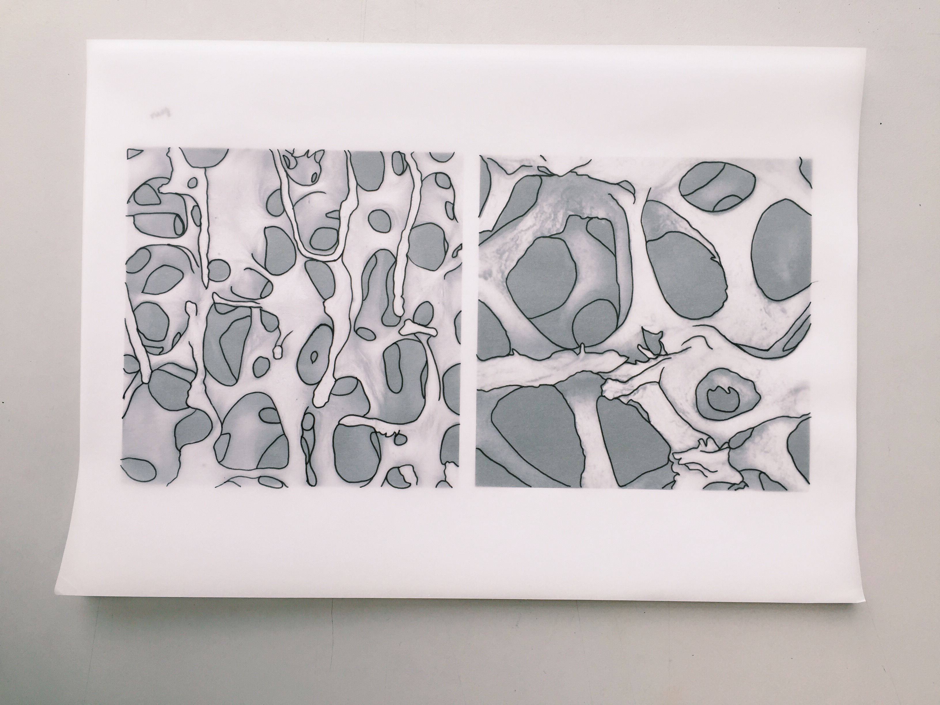
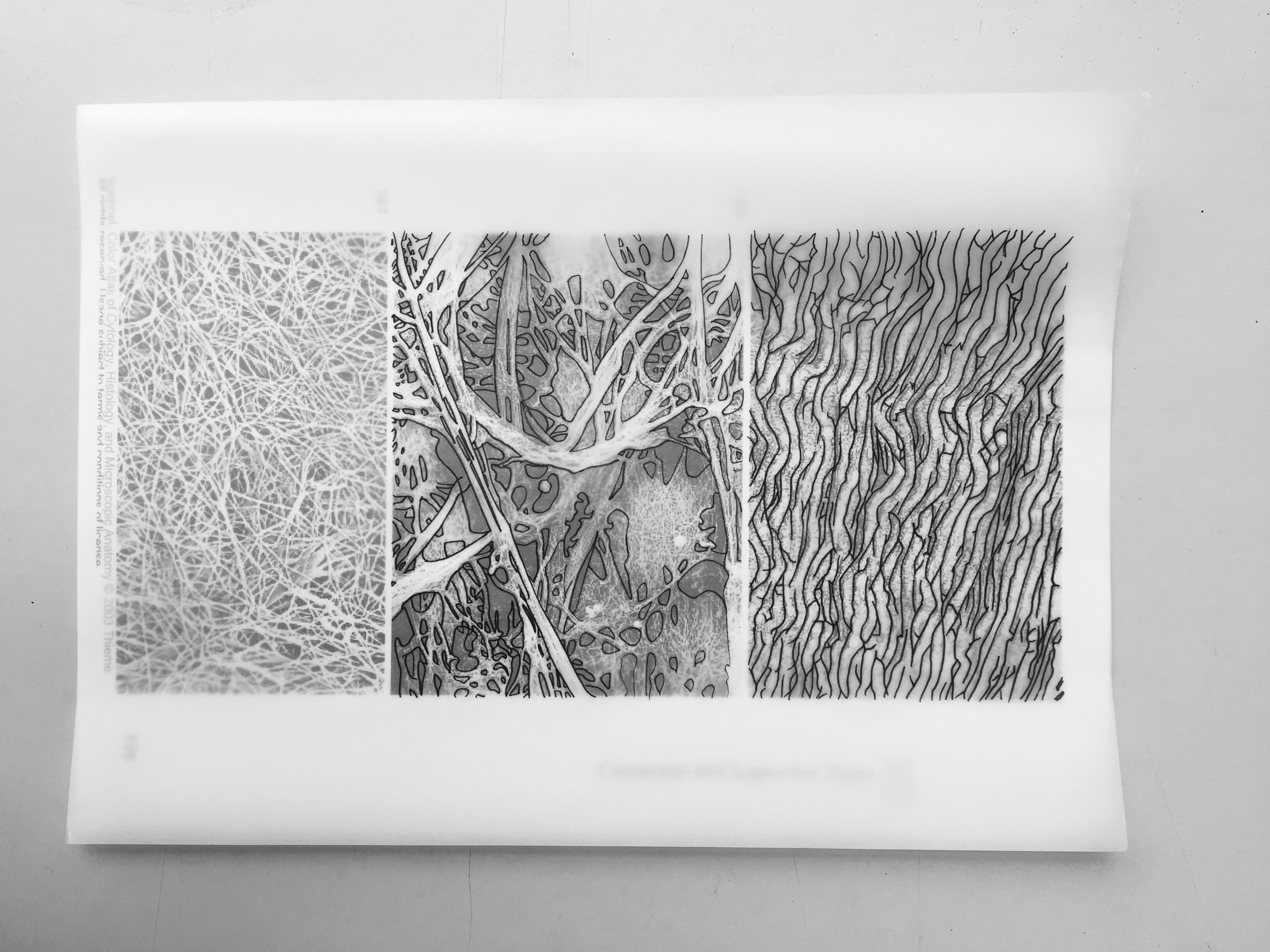
So from the previous post, I printed out selected images from a source that has data on human microscopic anatomy, traced those images, and lastly scanned them in. Not really at random, but I selected those images based on the type of patterns that I planned to incorporate into the entire motif — bones, cells, and connective and supportive tissues.
Softcopy
I did a few trial and error to vectorize the images that I scanned, and tried on getting a few motifs as a start. First thing first, I started by using each microscopic anatomy into its own motif by repetition within its own elements. I might say these trial motifs were headed towards the direction of symmetrical and geometrical patterns.
(Majority of the motifs are similar to the previous ones. Basically it shows the variations if I’d rather choose those motifs with intersecting lines in the middle or minimal intersection?)
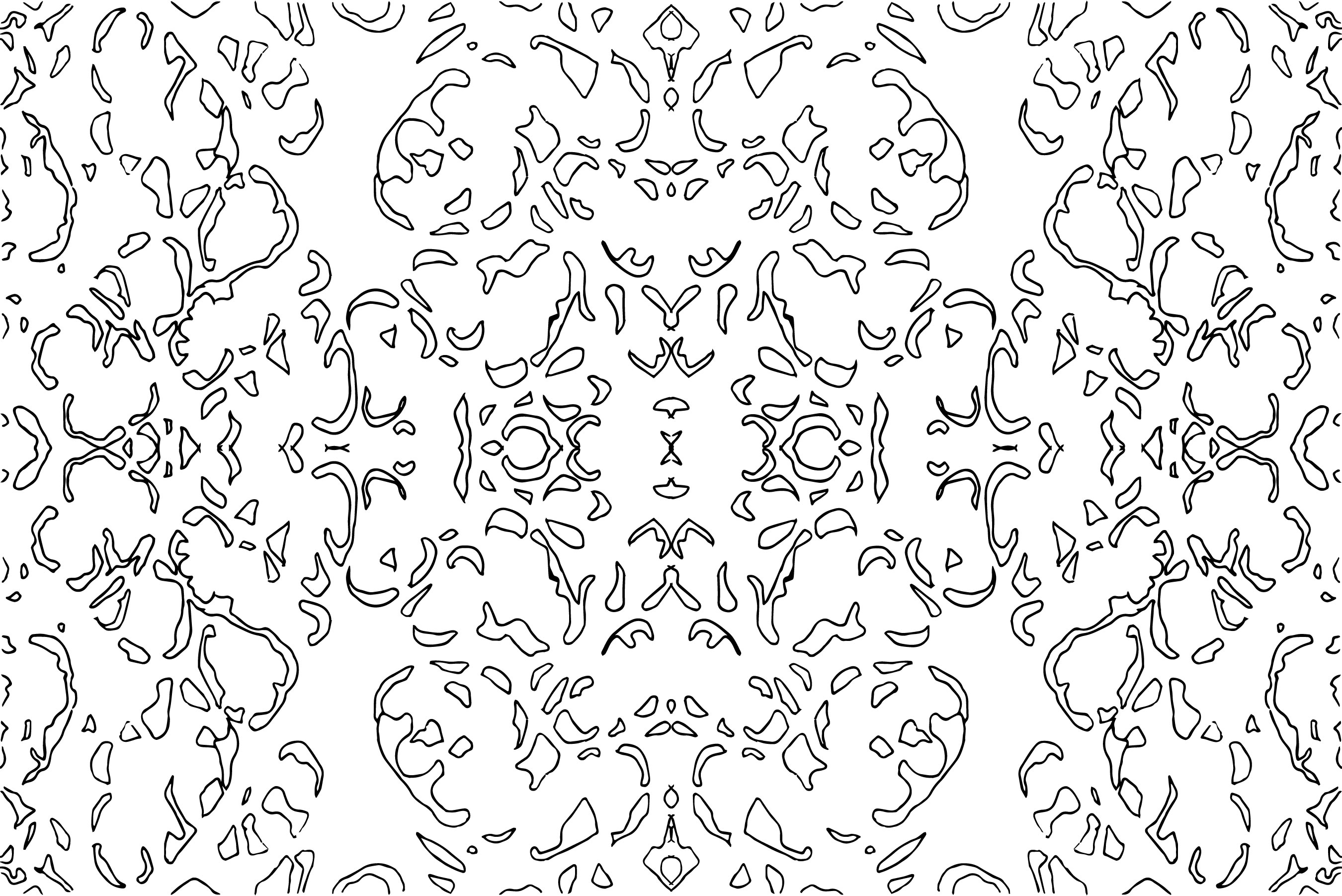
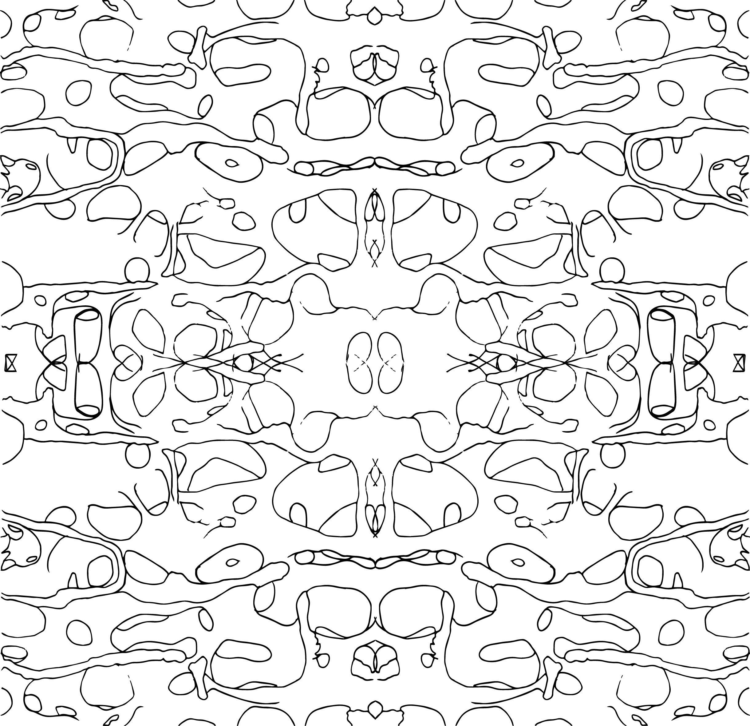
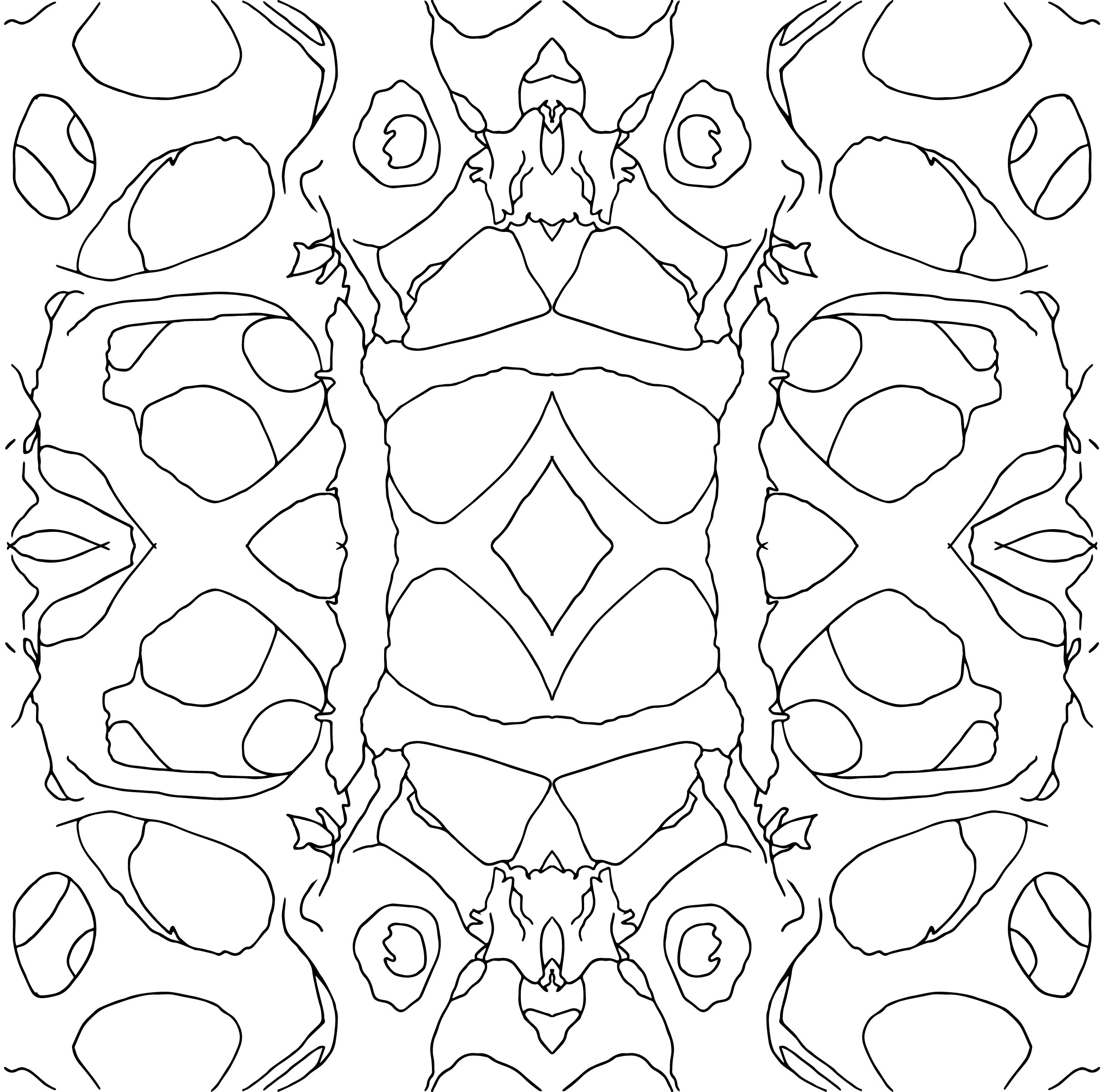
Motif #1: the microscopic structure of our bone #1.
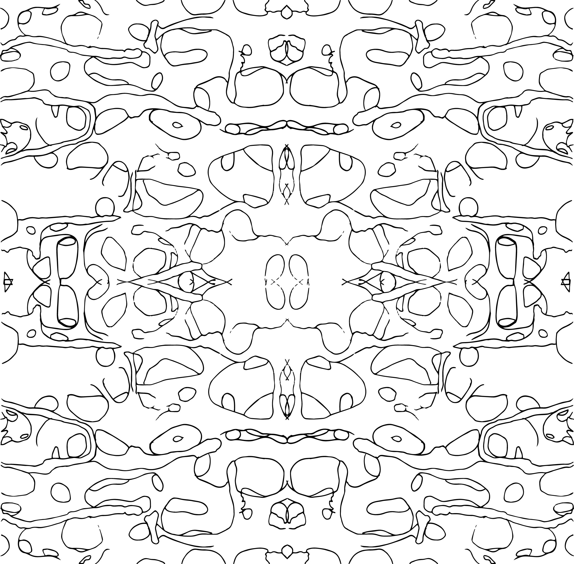
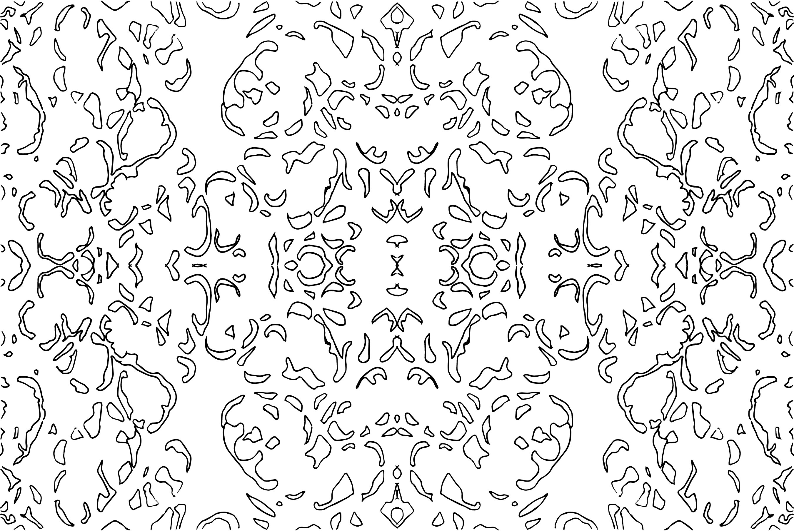
Motif #2: the microscopic structure of our bone #2.

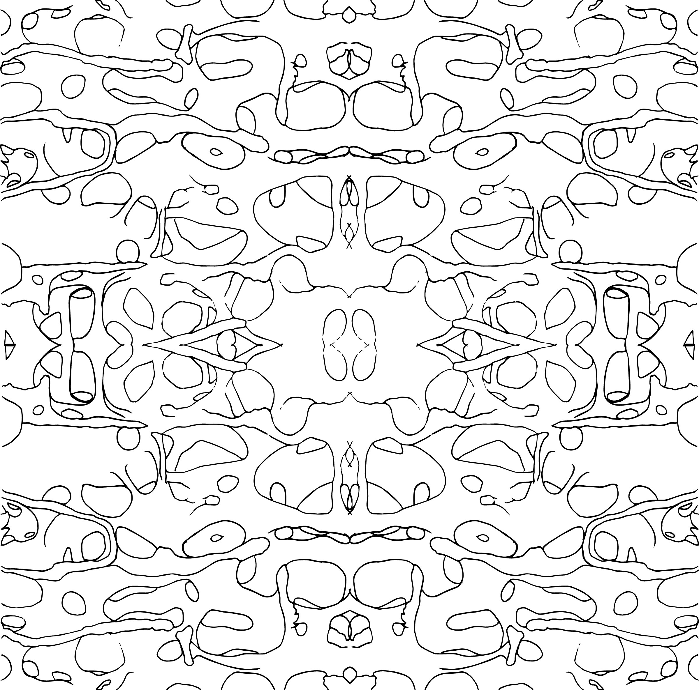
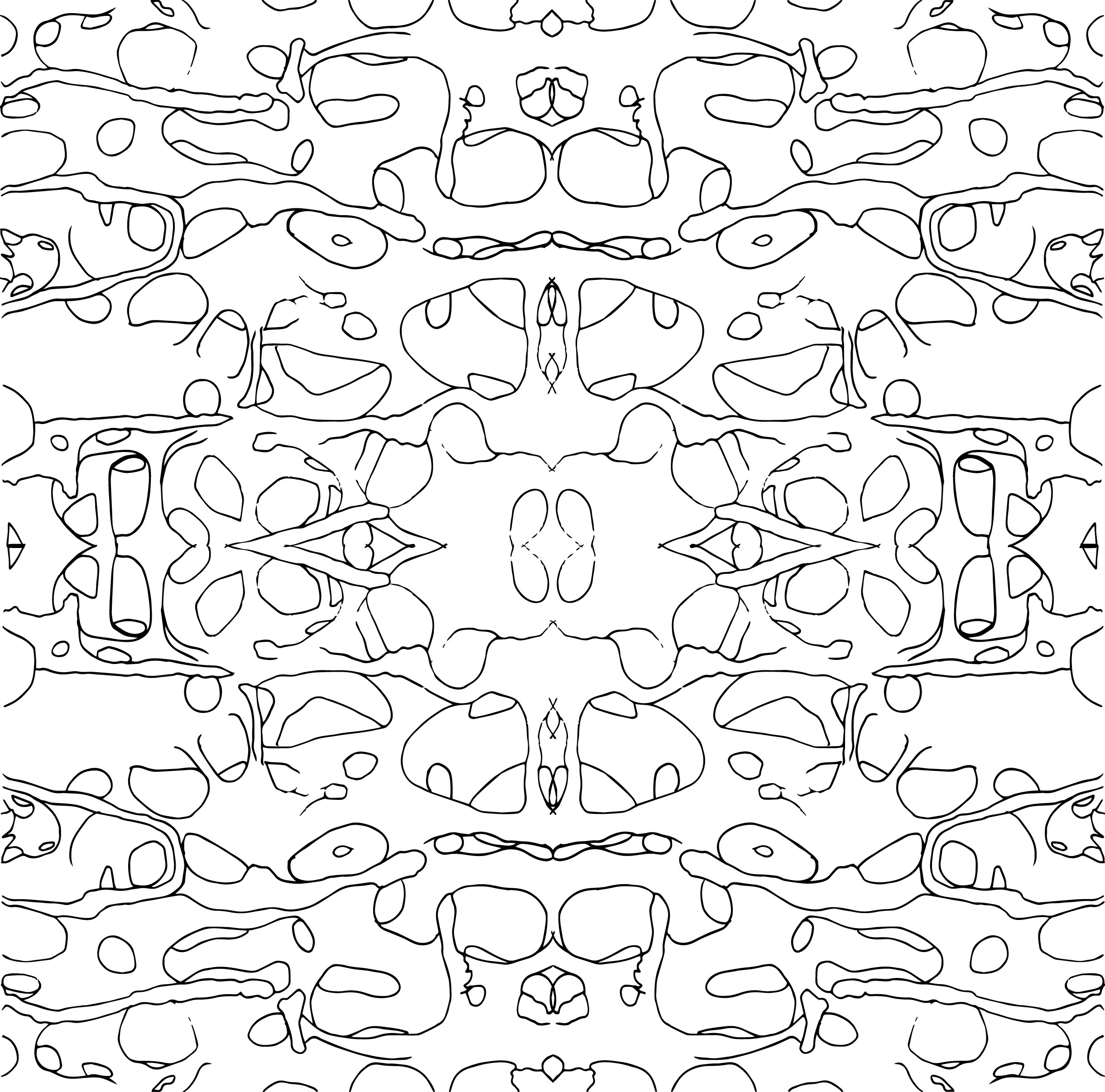
Motif #3: the microscopic structure of Connective and Supportive Tissue #1.

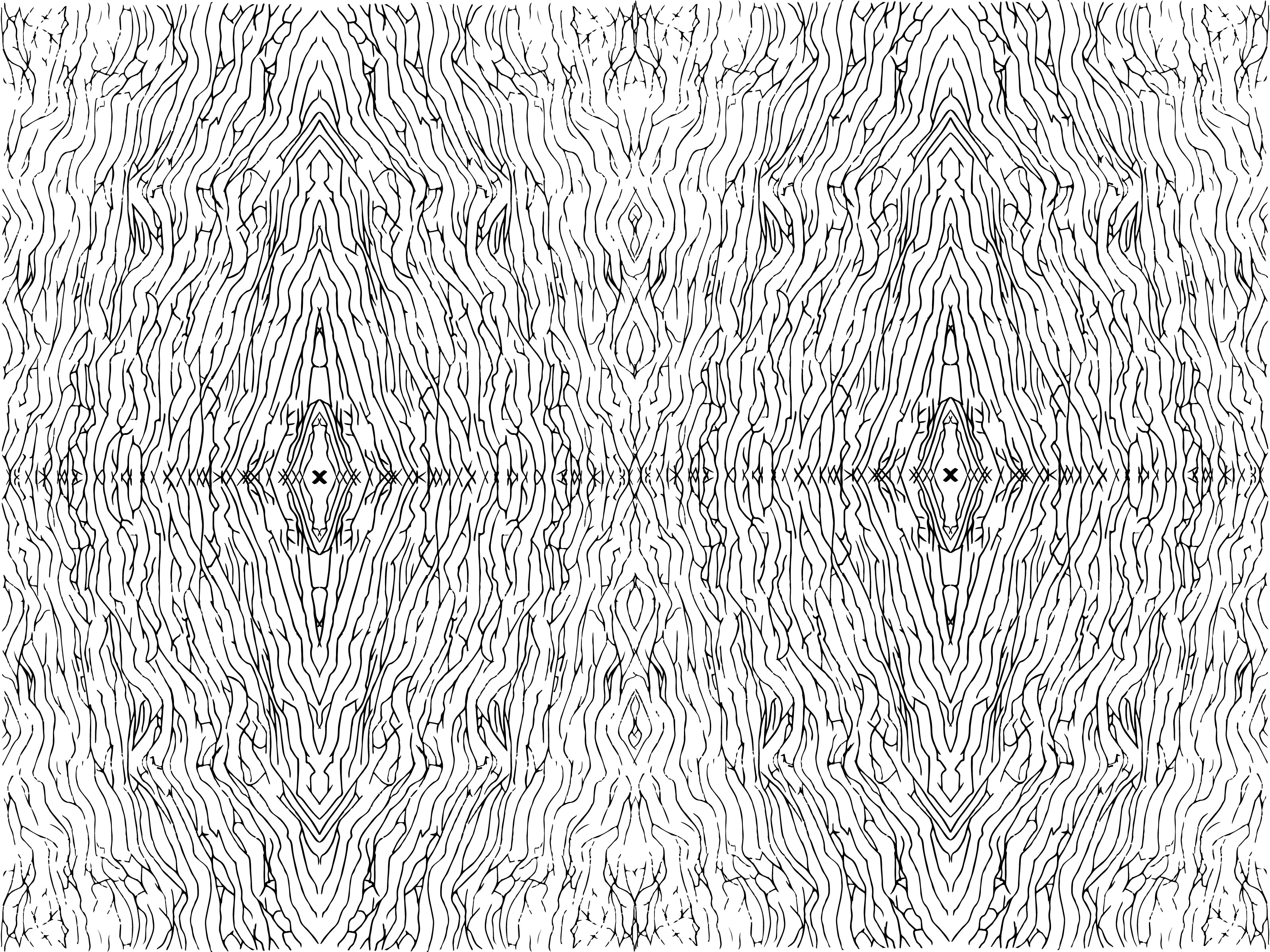
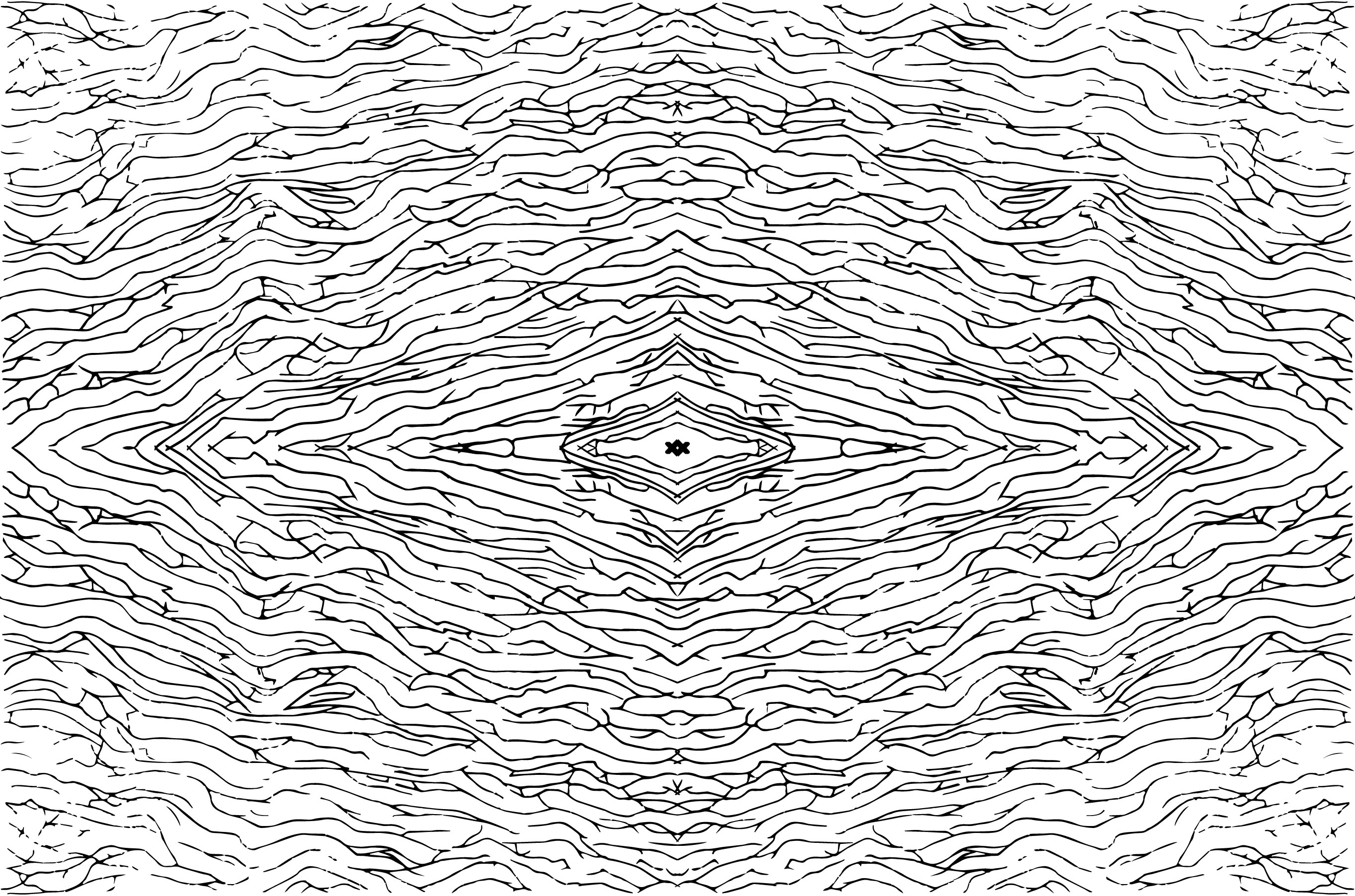
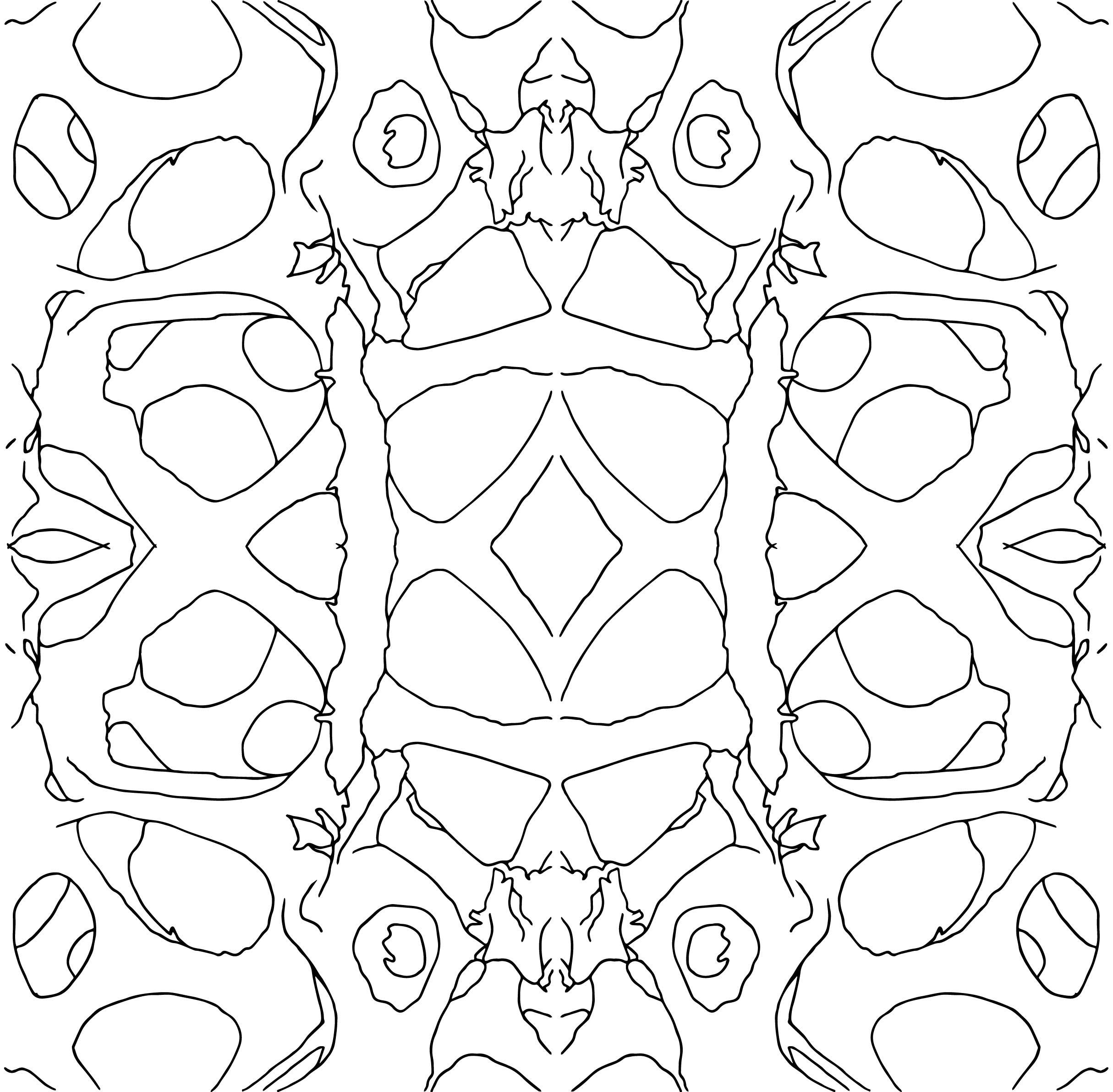
Motif #4: the microscopic structure of Connective and Supportive Tissue #2.
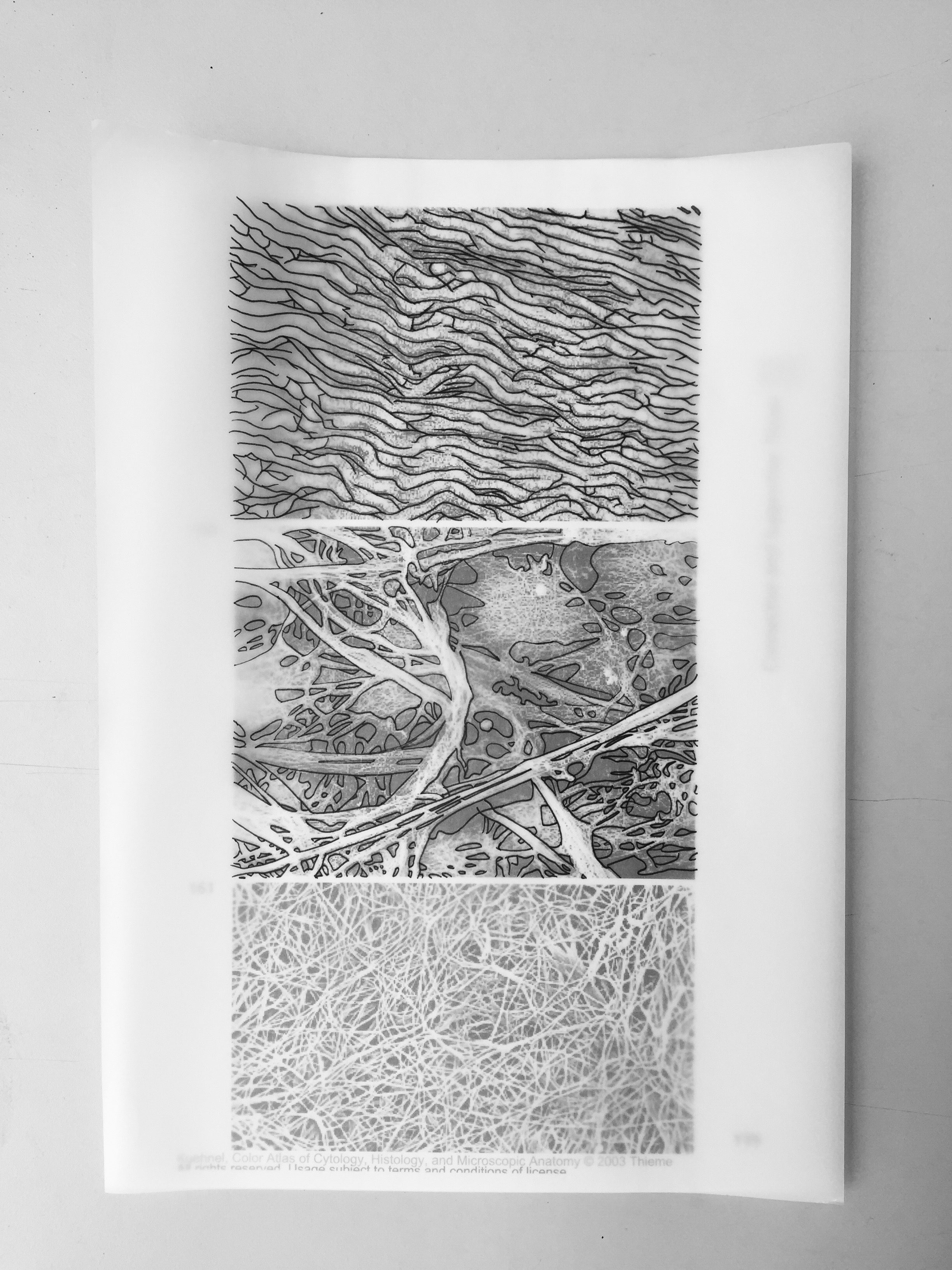
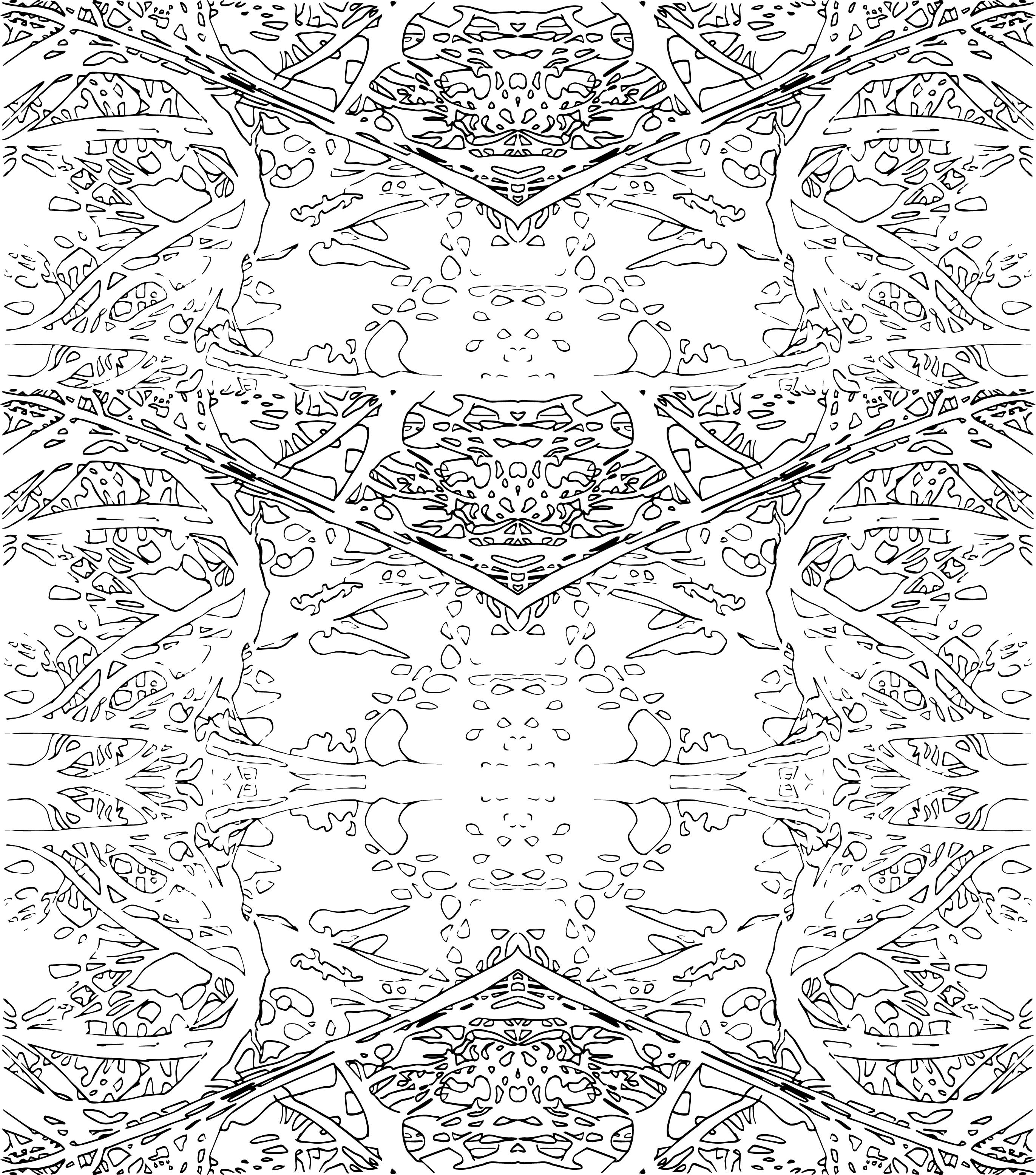
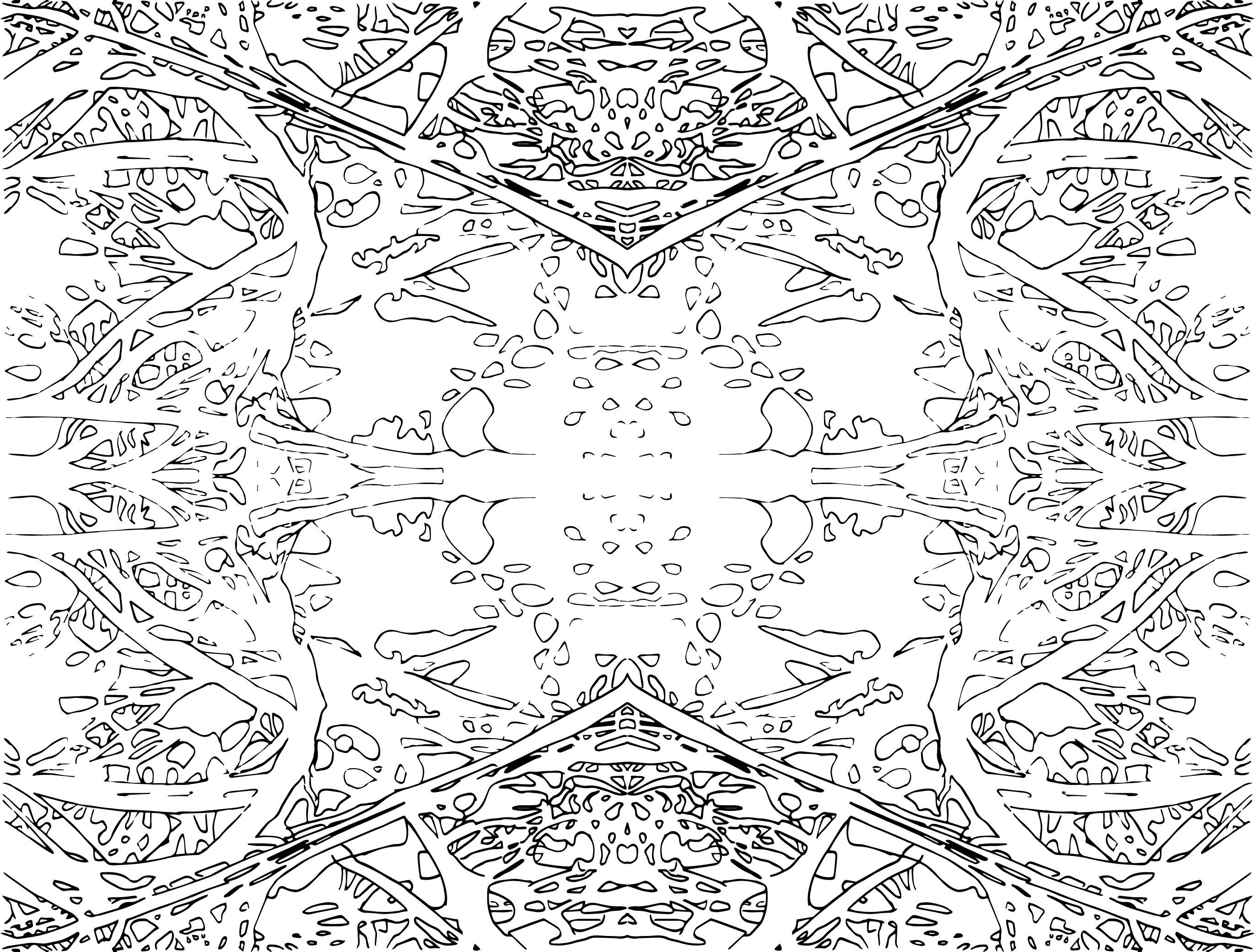
Motif #5: the microscopic structure of Connective and Supportive Tissue #3.


Motif #6: the microscopic structure of cells
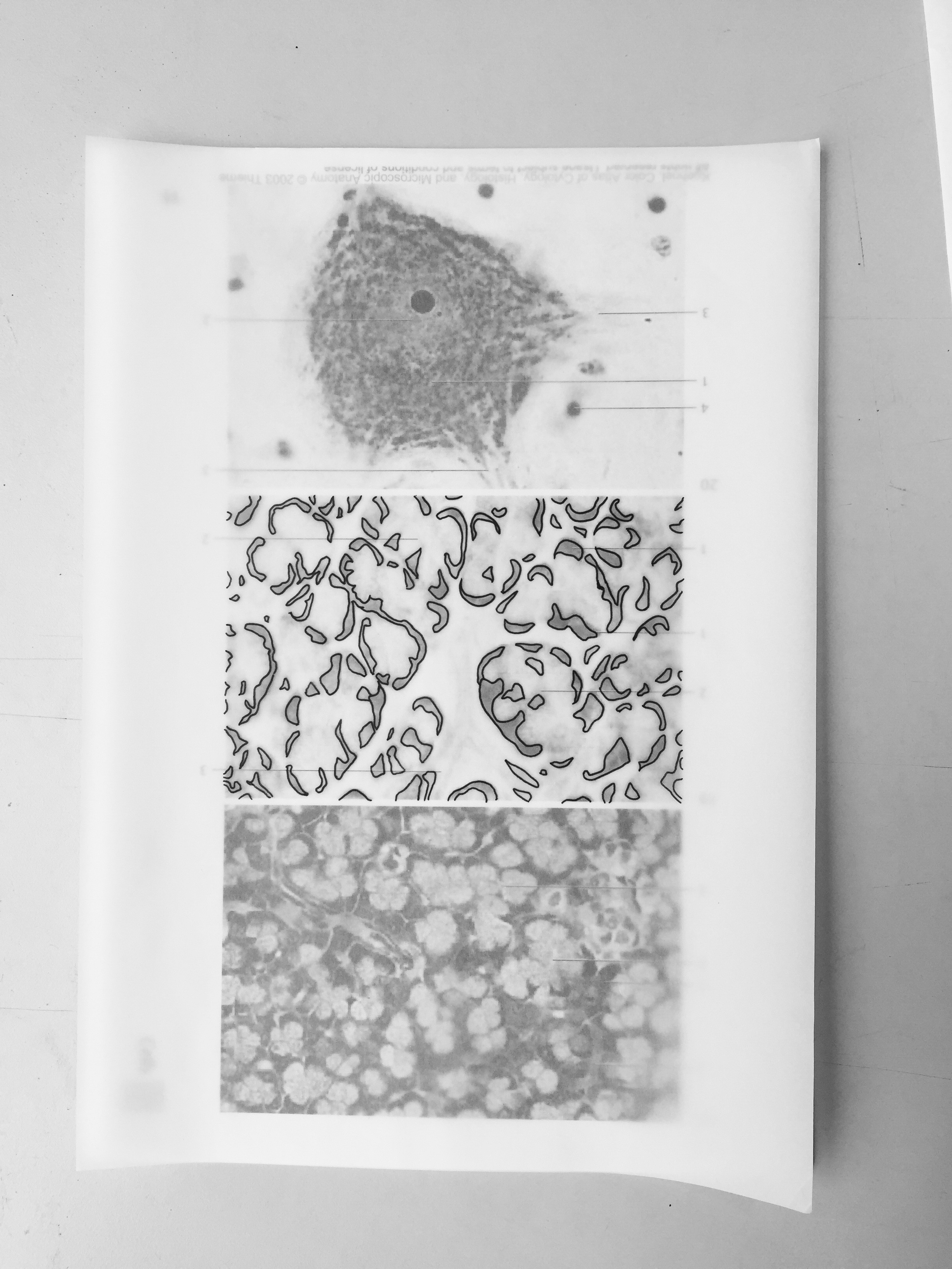
Kuehnel, Wolfgang. Color Atlas of Cytology, Histology, and Microscopic Anatomy. 4th ed. Germany: Thieme, 2003. PDF. Cells, pg 15